Date Milestones
Release Date
February 22, 2024
Review Date
Expiration Date
February 22, 2027
730 - Oral Diagnosis
February 22, 2024
February 22, 2027


Special Needs or Requests? Please Call the office for assistance.
CBCT, Oral Diagnosis, Radiology
The first module of this series “CBCT Foundations” is designed for those new to CBCT, those just getting started with their new Cone Beam CT system, and also as a refresher or reference tool for experienced CBCT users. In these videos, BeamReaders Oral and Maxillofacial Radiologist Dr. Kaycee Walton walks the viewer through basic definitions and concepts of CBCT usage.
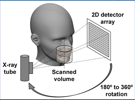
CBCT Foundations – Operating Principles: This video is the first in the CBCT Foundations series, by BeamReaders Oral and Maxillofacial Radiologist Dr. Kaycee Walton. Dr. Walton covers clinical considerations, hardware and the acquisition phase, software and the reconstruction phase, DICOM file export basics and best practices, and strengths and limitations of CBCT. General topics include definitions and practical application of raw data frames, fields of view, scan time, voxel size, milliampere settings, etc.
CBCT Foundations – Image Quality: This video is the second in the CBCT Foundations series, by BeamReaders Oral and Maxillofacial Radiologist Dr. Kaycee Walton. Dr. Walton covers qualitative measures of image quality, noise and artifacts, acquisition parameters, and image manipulation topics.
CBCT Foundations – Understanding Radiation Dose: This video is the third and final video in the CBCT Foundations series, by BeamReaders Oral and Maxillofacial Radiologist Dr. Kaycee Walton. Dr. Walton discusses the justification and optimization of radiation dose for patients, the biological effects of radiation, natural background radiation, factors influencing CBCT radiation dose, discussing the risk with patients, handling imaging during pregnancy, and appropriate shielding.
The second module of this series: Understanding 3D Anatomy is designed for dental professionals looking to review anatomy, and also see this anatomy as visualized by CBCT. These videos should both review and build upon previous anatomy learnings. In these videos, BeamReaders Oral and Maxillofacial Radiologist Dr. Shiva Toghyani walks the viewer through normal anatomy, structure, anatomical relation and as appropriate, common pathologies. There are a total of seven (7) topics in the complete series.
Sinuses: The video by BeamReaders Oral and Maxillofacial Radiologist Dr. Shiva Toghyani covers normal sinus anatomy, structure, anatomical relation and drainage pathways, and most common pathologies.
Airway: The video by BeamReaders Oral and Maxillofacial Radiologist Dr. Shiva Toghyani covers the upper airway anatomy, the sleep disordered breathing spectrum, and CBCT imaging of the airway.
TMJ: The video by BeamReaders Oral and Maxillofacial Radiologist Dr. Shiva Toghyani covers the temporomandibular joint anatomy, CBCT imaging of the TMJ, and the most common TMJ disorders.
Spine: The video by BeamReaders Oral and Maxillofacial Radiologist Dr. Shiva Toghyani covers normal cervical spine (C-spine) anatomy, CBCT imaging of the C-spine, and the most common C-spine abnormalities.
Soft Tissue Calcifications: This video by BeamReaders Oral and Maxillofacial Radiologist Dr. Martina Parrone will review common and uncommon soft tissue calcifications.
Nasal Cavity: The video by BeamReaders Oral and Maxillofacial Radiologist Dr. Martina Parrone covers normal nose and nasal cavity anatomy, a review of a proper search pattern for the nasal cavity, and common pathosis.
Nasal Cavity Bonus video: The video by BeamReaders Oral and Maxillofacial Radiologist Dr. Martina Parrone covers nasal cavity anatomical variants and common findings. These may be helpful since many of these variants are easy to confuse with to pathosis, and they can also play a role in sleep disordered breathing.
Dentition – Caries: The video by BeamReaders Oral and Maxillofacial Radiologist Dr. Martina Parrone will review dental caries as compared to a beam hardening artifact, which look very similar on a CBCT. She uses a case to share a cautionary tale to compare these two situations.
Dentition – Periodontal Disease: The video by BeamReaders Oral and Maxillofacial Radiologist Dr. Martina Parrone will review what periodontal disease looks like on a CBCT, both vertical defects and generalized horizontal bone loss, which look very similar on a CBCT. She uses a case to share how different views can provide insights to this evaluation.
Dentition – Periapical Pathosis & Endodontic Evaluation: The video series by BeamReaders Oral and Maxillofacial Radiologist Dr. Martina Parrone will use a case based approach to share what periapical lesions look like on CBCT, radiographic features that help distinguish inflammation from malignancy, and avoiding slice artifact. Cases compare unfilled canal spaces versus beam hardening artifacts, evaluating resorption, and reviewing periapical osseous dysplasia and idiopathic osteosclerosis.
The third and final module of this series: CBCT Search Pattern videos are designed for those new to CBCT. Experienced CBCT users will also gain valuable diagnostic pearls and learn about best practices when reviewing as CBCT scan! In these videos, BeamReaders Oral and Maxillofacial Radiologist Dr. Martina Parrone walks the viewer through a scan and findings. They are designed as a series, and intended to be watched sequentially. After initially viewing the entire sequence of videos, they may be used as a reference tool to review specific aspects of a scan and appropriate search pattern.
CBCT Intro, Orientation, Adjust Contrast, and Pano: These videos are the start of the CBCT Search Pattern series by BeamReaders Oral and Maxillofacial Radiologist Dr. Martina Parrone. These videos cover the technical analysis of a scan, orienting the scan in 3D, contrast adjustment, creating a panoramic image, and evaluating for asymmetries.
CBCT Evaluate Extraoral: These videos are the second part of the CBCT Search Pattern series by BeamReaders Oral and Maxillofacial Radiologist Dr. Martina Parrone. These videos cover the evaluation of extraoral structures in the sagittal, coronal, and axial planes.
CBCT Evaluate TMJ: This video is the third part of the CBCT Search Pattern series by BeamReaders Oral and Maxillofacial Radiologist Dr. Martina Parrone. This video covers the evaluation of the temporomandibular joint.
CBCT Dentition: These videos are the fourth part of the CBCT Search Pattern series by BeamReaders Oral and Maxillofacial Radiologist Dr. Martina Parrone. These videos cover the evaluation of dentoalveolar findings in the sagittal, coronal, and axial planes.
At the completion of this course the participants should be able to:
Special Needs or Requests? Please Call the office for assistance.
You are registering {{attendees.length}} attendees. You can add their info later.