

Presented by
John A. Svirsky
D.D.S., M.Ed.
View Bio
Course Description
Oral Pathology
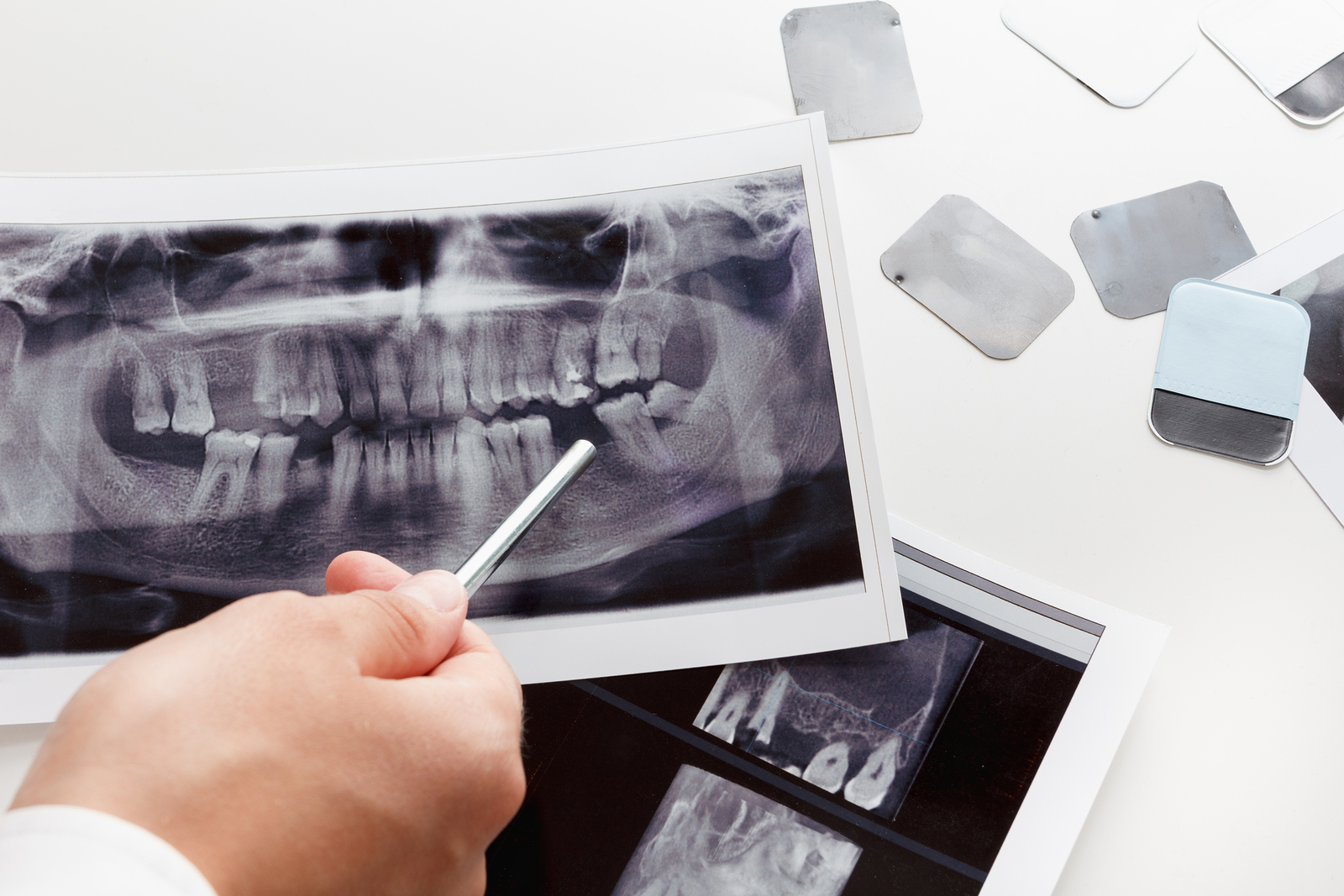
This live webinar is designed to be a full review of radiology for Dentists and Dental Hygienists. Dr. Svirsky will walk the audience through a number of common cases as a way of reacquianting the audience with key radiographic entities common to the dental practice. There will be an emphasis on common radiolucent and radiopaque lesions. In addition, Dr. Svirsky will spend some time highlighting the bizarre and unusual cases he’s encountered throughout his career.
By the end of this course, attendees will be well equipped to go back and make a difference in the diagnosis and treatment of oral diseases. This is the perfect course for dental professionals wanting a thorough review of radiology – everything you’ve learned in school, brought to you in shades of grey!
- Common/Important Radiolucencies
- Periapical Pathosis (cyst, granuloma, abscess, scar)
- Dentigerous Cyst
- Residual Cyst
- Odontogenic Keratocyst
- Traumatic Bone Cyst
- Lateral Periodontal Cyst
- Nasopalatine Duct Cyst
- Central Giant Cell Granuloma
- Ameloblastoma
- Lingual Mandibular Bone Defect
- Common/Important Radiopacities
- Condensing Osteitis
- Osteosclerosis
- Impacted Tooth
- Root Tips
- Odontoma
- Supernumerary Tooth
- Sialolth
- Common/Important Radiolucencies/Radiopacities
- Osteomyelitis
- Periapical Cemental Dysplasia
- Florid Osseous Dysplasia
- Central Ossifying Fibroma
Course Objectives
At the completion of this course the participants should be able to:
- Demonstrate a logical approach to the diagnosis and treatment of common radiolucent lesions found on radiographs.
- Demonstrate a logical approach to the diagnosis and treatment of common radiopaque lesions found on radiographs.
- Recognize the common radiographic lesion found in dental practices.

