Continuing Dental Education
If you prefer in-person seminars, live webinars, or on demand courses, we’ve got you covered. Please click on the options below to learn more!
Smart Savings on Live CE Webinars: Get $10 Off Per Webinar When You Purchase 2 or More at the Same Time!
 Seminar
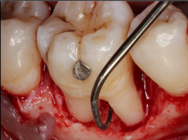
Clinical Periodontology for General Dentists and Dental Hygienists
8 Credit Hours
Friday Apr 26th, 2024
at 8:30 AM
The course is designed to discuss the contemporary aspects of clinical periodontology for general dentists and dental hygienists. Starting from the patient’s medical history and how important is to understand when to move forward with the data collection and in which circumstances to give a medical consultation. Participants will understand the etiological factors, the proper diagnosis and rationale behind the new classification system involving the staging and grading of chronic periodontitis.
Speaker: Nikolaos K. Soldatos
17 seats available
Indianapolis, IN
Seminar
Clinical Periodontology for General Dentists and Dental Hygienists
8 Credit Hours
Saturday Apr 27th, 2024
at 8:30 AM
The course is designed to discuss the contemporary aspects of clinical periodontology for general dentists and dental hygienists. Starting from the patient’s medical history and how important is to understand when to move forward with the data collection and in which circumstances to give a medical consultation. Participants will understand the etiological factors, the proper diagnosis and rationale behind the new classification system involving the staging and grading of chronic periodontitis.
Speaker: Nikolaos K. Soldatos
3 seats available
Fort Wayne, IN
 Seminar
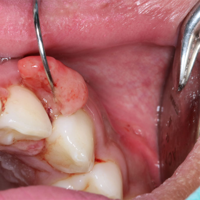
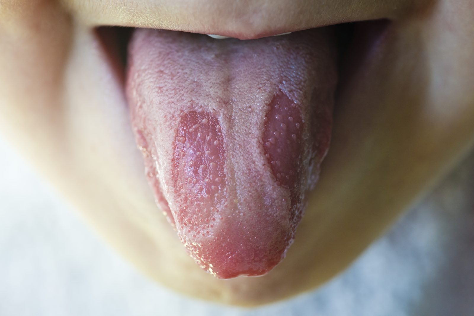
Oral Cancer & Diseases: Case Presentation of Lumps, Bumps & Lesions
8 Credit Hours
Friday Jul 12th, 2024
at 8:30 AM
This course deals with unusual cases, diagnostic challenges, the oral health effects of tobacco products and alcohol, premalignant and malignant lesions, presenting an oral cancer diagnosis to a patient and new diagnostic technique. Tobacco advertising will be exposed and the link between human papilloma virus and oral cancer discussed. Treatment of oral cancer and oropharyngeal cancer will be presented.
This course will be presented in Dr. Svirsky’s interactive and entertaining style. Get ready to laugh, learn and make a major difference in the lives of your patients.
Speaker: John A. Svirsky
97 seats available
Syracuse, NY
Seminar
Oral Cancer & Diseases: Case Presentation of Lumps, Bumps & Lesions
8 Credit Hours
Saturday Jul 13th, 2024
at 8:30 AM
This course deals with unusual cases, diagnostic challenges, the oral health effects of tobacco products and alcohol, premalignant and malignant lesions, presenting an oral cancer diagnosis to a patient and new diagnostic technique. Tobacco advertising will be exposed and the link between human papilloma virus and oral cancer discussed. Treatment of oral cancer and oropharyngeal cancer will be presented.
This course will be presented in Dr. Svirsky’s interactive and entertaining style. Get ready to laugh, learn and make a major difference in the lives of your patients.
Speaker: John A. Svirsky
149 seats available
Saratoga Springs, NY
 Seminar
Non-Surgical Periodontal Therapy: Disease Identification, Instrumentation, and Evidence-Based Updates
8 Credit Hours
Friday Jul 26th, 2024
at 8:30 AM
This course is designed for dentists & dental hygienists and will offer information to identify signs and symptoms of periodontal disease. Dr. Sharma will review advanced instrumentation to avoid burnishing and effectively remove calculus during non-surgical periodontal therapy and the most current research and guidelines related to NSPT, providing valuable insight into effectively treating and stabilizing periodontal disease.
Speaker: Silky Sharma
100 seats available
Jessup, MD
Seminar
Non-Surgical Periodontal Therapy: Disease Identification, Instrumentation, and Evidence-Based Updates
8 Credit Hours
Saturday Jul 27th, 2024
at 8:30 AM
This course is designed for dentists & dental hygienists and will offer information to identify signs and symptoms of periodontal disease. Dr. Sharma will review advanced instrumentation to avoid burnishing and effectively remove calculus during non-surgical periodontal therapy and the most current research and guidelines related to NSPT, providing valuable insight into effectively treating and stabilizing periodontal disease.
Speaker: Silky Sharma
99 seats available
Harrisburg, PA
Seminar
Non-Surgical Periodontal Therapy: Disease Identification, Instrumentation, and Evidence-Based Updates
8 Credit Hours
Friday Aug 9th, 2024
at 8:30 AM
This course is designed for dentists & dental hygienists and will offer information to identify signs and symptoms of periodontal disease. Dr. Sharma will review advanced instrumentation to avoid burnishing and effectively remove calculus during non-surgical periodontal therapy and the most current research and guidelines related to NSPT, providing valuable insight into effectively treating and stabilizing periodontal disease.
Speaker: Silky Sharma
100 seats available
Plymouth Meeting, PA
Seminar
Oral Cancer & Diseases: Case Presentation of Lumps, Bumps & Lesions
8 Credit Hours
Friday Aug 9th, 2024
at 8:30 AM
This course deals with unusual cases, diagnostic challenges, the oral health effects of tobacco products and alcohol, premalignant and malignant lesions, presenting an oral cancer diagnosis to a patient and new diagnostic technique. Tobacco advertising will be exposed and the link between human papilloma virus and oral cancer discussed. Treatment of oral cancer and oropharyngeal cancer will be presented.
This course will be presented in Dr. Svirsky’s interactive and entertaining style. Get ready to laugh, learn and make a major difference in the lives of your patients.
Speaker: John A. Svirsky
88 seats available
Live Oak, TX
Seminar
Non-Surgical Periodontal Therapy: Disease Identification, Instrumentation, and Evidence-Based Updates
8 Credit Hours
Saturday Aug 10th, 2024
at 8:30 AM
This course is designed for dentists & dental hygienists and will offer information to identify signs and symptoms of periodontal disease. Dr. Sharma will review advanced instrumentation to avoid burnishing and effectively remove calculus during non-surgical periodontal therapy and the most current research and guidelines related to NSPT, providing valuable insight into effectively treating and stabilizing periodontal disease.
Speaker: Silky Sharma
150 seats available
Lancaster, PA
Seminar
Oral Cancer & Diseases: Case Presentation of Lumps, Bumps & Lesions
8 Credit Hours
Saturday Aug 10th, 2024
at 8:30 AM
This course deals with unusual cases, diagnostic challenges, the oral health effects of tobacco products and alcohol, premalignant and malignant lesions, presenting an oral cancer diagnosis to a patient and new diagnostic technique. Tobacco advertising will be exposed and the link between human papilloma virus and oral cancer discussed. Treatment of oral cancer and oropharyngeal cancer will be presented.
This course will be presented in Dr. Svirsky’s interactive and entertaining style. Get ready to laugh, learn and make a major difference in the lives of your patients.
Speaker: John A. Svirsky
120 seats available
Austin, TX
 Seminar
Comprehensive Overview of Oral Pathology
8 Credit Hours
Friday Aug 16th, 2024
at 8:30 AM
This course will provide attendees with a comprehensive review of oral pathology. Common entities will be discussed; a particular focus will be made on oral squamous cell carcinoma and prevention of this malignancy.
Speaker: Ashley N. Clark
65 seats available
Santa Fe, NM
Seminar
Comprehensive Overview of Oral Pathology
8 Credit Hours
Saturday Aug 17th, 2024
at 8:30 AM
This course will provide attendees with a comprehensive review of oral pathology. Common entities will be discussed; a particular focus will be made on oral squamous cell carcinoma and prevention of this malignancy.
Speaker: Ashley N. Clark
130 seats available
Albuquerque, NM
 Seminar
Infection Control & Safety in the Dental Setting
8 Credit Hours
Friday Nov 15th, 2024
at 8:30 AM
As we begin to look at the COVID-19 pandemic in the rear-view mirror, the science and understanding and of this disease continues to evolve, and new emerging infectious diseases are now within our communities. While disease transmission in dentistry is rare, consequences may be severe. Participants will learn the latest policies and procedures that impact effective infection control in the dental setting. And, the entire dental team will learn that infection prevention is a team-sport that requires constant attention, education, and compliance.
Speaker: Marie Fluent
130 seats available
Freeport, ME
Seminar
Infection Control & Safety in the Dental Setting
8 Credit Hours
Saturday Nov 16th, 2024
at 8:30 AM
As we begin to look at the COVID-19 pandemic in the rear-view mirror, the science and understanding and of this disease continues to evolve, and new emerging infectious diseases are now within our communities. While disease transmission in dentistry is rare, consequences may be severe. Participants will learn the latest policies and procedures that impact effective infection control in the dental setting. And, the entire dental team will learn that infection prevention is a team-sport that requires constant attention, education, and compliance.
Speaker: Marie Fluent
130 seats available
Manchester, NH
Seminar
Comprehensive Overview of Oral Pathology
8 Credit Hours
Friday Nov 22nd, 2024
at 8:30 AM
This course will provide attendees with a comprehensive review of oral pathology. Common entities will be discussed; a particular focus will be made on oral squamous cell carcinoma and prevention of this malignancy.
Speaker: Ashley N. Clark
174 seats available
Nashua, NH
Seminar
Comprehensive Overview of Oral Pathology
8 Credit Hours
Saturday Nov 23rd, 2024
at 8:30 AM
This course will provide attendees with a comprehensive review of oral pathology. Common entities will be discussed; a particular focus will be made on oral squamous cell carcinoma and prevention of this malignancy.
Speaker: Ashley N. Clark
148 seats available
Salem, MA
 Seminar
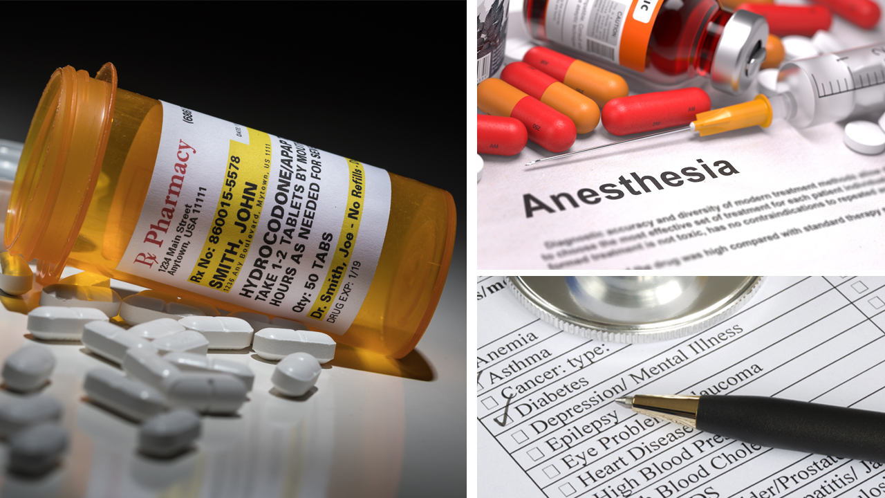
“It’s All About The Medical History!”: Essential Pharmacology for Busy Dental Professionals
8 Credit Hours
Friday Dec 6th, 2024
at 8:30 AM
What potential complications lurk behind the patient’s medical history that may impact dental therapy? This program will provide an overview of the dental implications of the medications, supplements and substances of abuse most frequently used by patients. The pharmacology of pain control and the effective management of acute dental pain using non-opioid and opioid analgesics will be presented as well as an overview of the pharmacology and therapeutics of local anesthetic agents.
Speaker: Thomas A. Viola
250 seats available
Feasterville-Trevose, PA
Seminar
“It’s All About The Medical History!”: Essential Pharmacology for Busy Dental Professionals
8 Credit Hours
Saturday Dec 7th, 2024
at 8:30 AM
What potential complications lurk behind the patient’s medical history that may impact dental therapy? This program will provide an overview of the dental implications of the medications, supplements and substances of abuse most frequently used by patients. The pharmacology of pain control and the effective management of acute dental pain using non-opioid and opioid analgesics will be presented as well as an overview of the pharmacology and therapeutics of local anesthetic agents.
Speaker: Thomas A. Viola
156 seats available
Hershey, PA
Seminar
“It’s All About The Medical History!”: Essential Pharmacology for Busy Dental Professionals
8 Credit Hours
Sunday Dec 8th, 2024
at 8:30 AM
What potential complications lurk behind the patient’s medical history that may impact dental therapy? This program will provide an overview of the dental implications of the medications, supplements and substances of abuse most frequently used by patients. The pharmacology of pain control and the effective management of acute dental pain using non-opioid and opioid analgesics will be presented as well as an overview of the pharmacology and therapeutics of local anesthetic agents.
Speaker: Thomas A. Viola
395 seats available
Cherry Hill, NJ